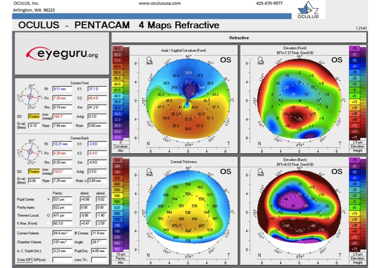
Placido Disc Uses / Placido Disc And Representative Patterns Of Corneal Shapes Download Scientific Diagram : In the final instalment of our series on taking good topography maps, we will look at the importance of capturing topography maps with good corneal coverage.
Placido Disc Uses / Placido Disc And Representative Patterns Of Corneal Shapes Download Scientific Diagram : In the final instalment of our series on taking good topography maps, we will look at the importance of capturing topography maps with good corneal coverage.. Rs 1,800 / piece get latest price. After projecting a concentric annular light source onto the corneal surface, placido disc reflection systems capture the reflected light so their software can measure curvature, irregularities, foreign bodies, tear film nuances and other characteristics of the anterior cornea. © 2003 by saunders, an imprint of elsevier, inc. The original style topographers use placido disc technology. A plurality of opaque, concentric rings are formed on the substrate.
Z2 general categories {curvature based systems zsystems that employ a large placido disk which is several inches in diameter and is positioned several inches from the patient's eye where the imaging is performed zsystems that employ small placido cone disk that fit very close to the eye when the imaging is performed. The substrate is configured to be activated by electric current and responsive to emit light. A keratoscope, sometimes known as placido's disk, is an ophthalmic instrument used to assess the shape of the anterior surface of the cornea. Corneal topography is a procedure used to monitor and measure changes that may occur to the shape and integrity of the cornea of your eye. Placido disc evaluation can also be used to guide suture removal following penetrating keratoplasty.

To realize the precise detection of corneal surface topography, an optical system for the corneal topography that is based on a placido disc is designed, which includes a ring distribution on a placido disc, an imaging system and a.
You can change your ad preferences anytime. A placido projector for a corneal topography system includes a substrate having dielectric phosphor. A placido disk is projected onto the cornea and the images of the placido disk reflected off the cornea are captured. Z2 general categories {curvature based systems zsystems that employ a large placido disk which is several inches in diameter and is positioned several inches from the patient's eye where the imaging is performed zsystems that employ small placido cone disk that fit very close to the eye when the imaging is performed. Corneal topography is a procedure used to monitor and measure changes that may occur to the shape and integrity of the cornea of your eye. Rs 1,800 / piece get latest price. Cataract surgery • preoperative use: The original style topographers use placido disc technology. In the final instalment of our series on taking good topography maps, we will look at the importance of capturing topography maps with good corneal coverage. A keratoscope, sometimes known as placido's disk, is an ophthalmic instrument used to assess the shape of the anterior surface of the cornea. A series of concentric rings is projected onto the. Corneal topography provides powerful support in the diagnosis and treatment of corneal disease by displaying the corneal surface topography in data or image format. When the substrate emits light responsive to the electric current, a placido image is projected.
1) uveitis , 2) keratoconus , 3) retinoblastoma , 4) retinal detachment Placido disc, clip, and handle this economical keratoscope is used to determine the curvature characteristics of the anterior surface of the cornea. A series of concentric rings is projected onto the cornea and their reflection viewed by the examiner through a small hole in the centre of the disk. Corneal topography provides powerful support in the diagnosis and treatment of corneal disease by displaying the corneal surface topography in data or image format. Rigid gas permeable contact lenses are needed in the majority of cases to neutralize the irregular corneal astigmatism.

This is the principle used for purkinje imaging as well in the placido discs.
A series of concentric rings is projected onto the cornea and their reflection viewed by the examiner through a small hole in the centre of the disk. A placido projector for a corneal topography system includes a substrate having dielectric phosphor. 1) uveitis , 2) keratoconus , 3) retinoblastoma , 4) retinal detachment This is the principle used for purkinje imaging as well in the placido discs. A keratoscope, sometimes known as placido's disk, is an ophthalmic instrument used to assess the shape of the anterior surface of the cornea. It is the evaluation of the corneal surface using circular mires reflected from its surface. However, an absolute height map would not tell us much other than that the cornea is a sphere, which is not a real surprise. A keratoscope, sometimes known as placido's disk, is an ophthalmic instrument used to assess the shape of the anterior surface of the cornea. A placido disk is projected onto the cornea and the images of the placido disk reflected off the cornea are captured. Placido disc systems use the tangential data to generate height maps. The original style topographers use placido disc technology. To realize the precise detection of corneal surface topography, an optical system for the corneal topography that is based on a placido disc is designed, which includes a ring distribution on a placido disc, an imaging system and a. The substrate is configured to be activated by electric current and responsive to emit light.
Z2 general categories {curvature based systems zsystems that employ a large placido disk which is several inches in diameter and is positioned several inches from the patient's eye where the imaging is performed zsystems that employ small placido cone disk that fit very close to the eye when the imaging is performed. Corneal topography provides powerful support in the diagnosis and treatment of corneal disease by displaying the corneal surface topography in data or image format. 1) uveitis , 2) keratoconus , 3) retinoblastoma , 4) retinal detachment This is the principle used for purkinje imaging as well in the placido discs. A placido disk is projected onto the cornea and the images of the placido disk reflected off the cornea are captured.

This code constructs curvature topography from placido rings image.
1) uveitis , 2) keratoconus , 3) retinoblastoma , 4) retinal detachment After projecting a concentric annular light source onto the corneal surface, placido disc reflection systems capture the reflected light so their software can measure curvature, irregularities, foreign bodies, tear film nuances and other characteristics of the anterior cornea. Corneal topography is used to characterize the shape of the cornea, specifically, the anterior surface of the cornea. The earliest device designed to perform this function was the placido's disc, developed by antonio placido. During a cvak procedure, the patient is seated in front of a bowl that contains an illuminated pattern on a placido cone disk. A corneal topographer projects a series of illuminated rings, referred to as a placido disc, onto the surface of the cornea, which are reflected back into the instrument. Z2 general categories {curvature based systems zsystems that employ a large placido disk which is several inches in diameter and is positioned several inches from the patient's eye where the imaging is performed zsystems that employ small placido cone disk that fit very close to the eye when the imaging is performed. Placido disc systems use the tangential data to generate height maps. You can change your ad preferences anytime. Cataract surgery • preoperative use: A keratoscope, sometimes known as placido's disk, is an ophthalmic instrument used to assess the shape of the anterior surface of the cornea. It consists of equally spaced alternating black and white rings with a hole in the centre to observe the patient's cornea. Rs 1,800 / piece get latest price.
Komentar
Posting Komentar